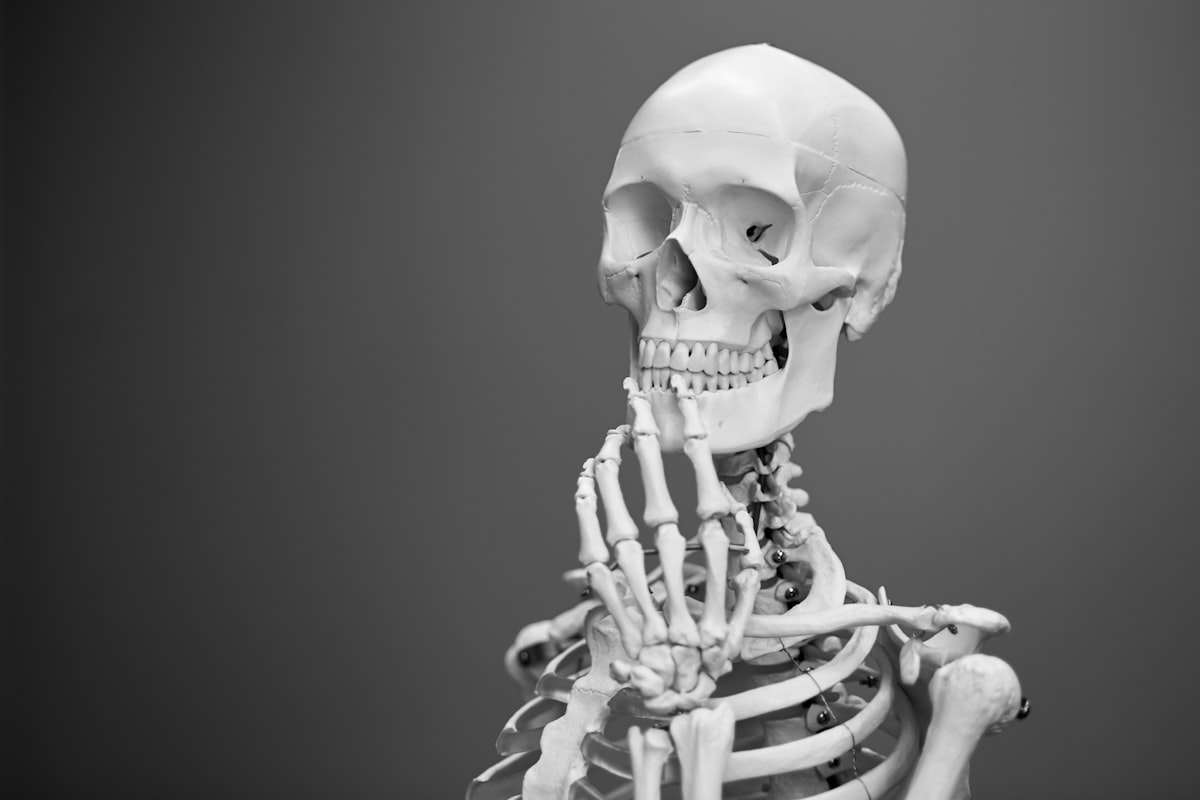
How the Orbit and Frontal Bones Are Connected to the Skull

There are many parts to a human skull, including the bones that form the orbit and frontal bone. This article will discuss how they are connected to the skull. You will also learn more about the sphenoid bone, the bony structure that forms the ear, and the bones that make up the cranium. It is not possible to know all of the parts of the skull, but this article will cover most of them.
Bones that make up the skull
The human skull is made up of 44 bones, many of which fuse together gradually to form a solid bone. In the initial stage, the bones of the roof of the skull are separated by regions of dense connective tissue called fontanelles. These organs allow the skull to develop during birth, but also allow it to expand in early infancy. As the skull grows, the fontanels are gradually removed and the bones fuse together as solid bone.
The skull is made up of 8 cranial bones, as well as the 14 facial bones that make up the nose, mouth, and jaw. The bones that make up the nose are small, and include the nasal bone and the conchae. The frontal bone forms the forehead, while the parietal bones are behind the frontal bone. The brain sits inside the cranium. A skull that protects the brain is essential to the brain's health.
Bones that form the walls of the orbit
The walls of the orbit are formed by four bones: the frontal bone, zygomatic bone, and the palatine bone. The frontal bone forms the superior and lateral margins of the orbit. The zygomatic bone forms the inferior and lateral walls of the orbit. The frontal bone also forms the supraorbital foramen, which transmits the orbital nerve and artery.
The bony orbits are cavities in the skull, located on either side of the forehead. These cavities house the eyeball and its associated structures. They form a pyramidal structure, with apex posteriorly and base anteriorly. The orbital walls are formed by seven bones. Their anatomical relationship is important for understanding how the eyeball and related structures are located in the skull.
Bones that form the frontal bone
The frontal bone in the skull is a complex piece of skull anatomy. It surrounds the brain in the cranial cavity, forming the upper and lower jaws and the nose and orbits. It also forms the nasal cavity. The frontal bone is rounded, and the pterion is the most prone to fracture. Other bones in the skull make up the skull's walls, including the temporal bone.
The lateral skull is dominated by the large rounded brain case, and the upper and lower jaws. The frontal bone is divided from the rest of the skull by the zygomatic arch, a narrow ridge formed by the zygomatic process of the temporal bone. The frontal bone, along with the parietal and occipital bones, form the frontal bone's inner surface.
Bones that form the sphenoid bone
The sphenoid bone is a single midline bone in the skull that articulates with the frontal, ethmoid, temporal, and palatine bones. It is located at the base of the skull, forming the floor and anterior walls of the middle cranial fossa. It contains multiple openings, including the optic canal and the superior orbital fissure.
The body of the sphenoid is a triangular-shaped structure that comprises the central part of the middle fossa, separating the chiasmal sulcus from the posterior clinoid process. The sphenoid bone is divided into three sections: the tuberculum sellae, a small olive-shaped swelling between the chiasmal sulcus and the posterior clinoid process, and the dorsum sellae, the furthest part. The latter contains a groove that serves as the exit point for the ICA, which passes through the petrous apex of the sphenoid bone.


