
Lymphatic Drainage of the Stomach
The lymphatic drainage of the stomach is a natural process that helps to eliminate excess fluid in the abdomen. The lymph nodes do it in the abdominal organs, the Submucosal plexus, Lesser omentum, and Greater omentum. Learn how to breathe properly to increase lymphatic flow.
Abdominal organs drain into abdominal lymph nodes.
Abdominal lymph nodes are the drainage systems for various internal organs, such as the liver, spleen, and pancreas. They drain into large sacks called cisterna chyle. The lymphatic vessels are thin and hard to see without a microscope, producing white blood cells called T lymphocytes and B lymphocytes. These cells carry large protein particles to the bloodstream. The lymphatic system also assists in maintaining blood volume.
The abdominal lymphatic system comprises four main groups of lymph nodes. The anterior group, known as the preaortic lymph nodes, is anterior to the abdominal aorta and consists of the celiac, inferior, and mesenteric lymph nodes. Each group drains organs supplied by similar arteries.
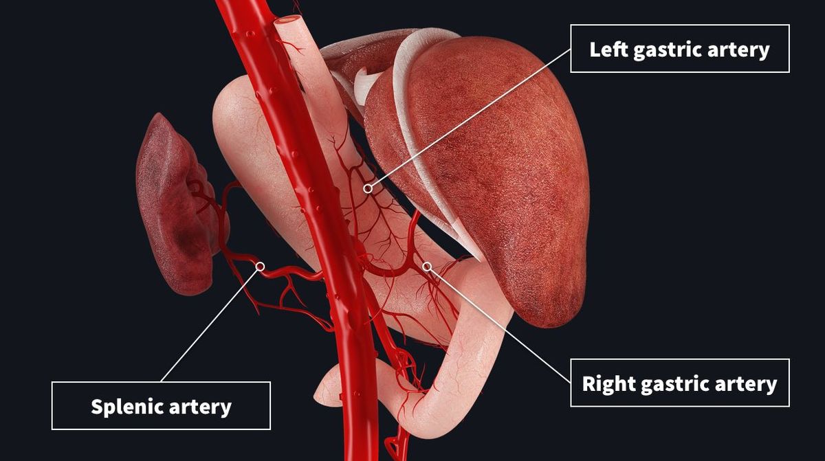
Greater omentum
The greater omentum is an area of the stomach with lymphatic connections to several other organs. It is also a well-known site for abdominal cancer metastasis. Because of its lymphatic connections, the greater omentum has been studied better to understand the lymphatic system's function in this area. This research involved 10 mongrel dogs anesthetized using veterinary pentobarbital sodium, endotractutubation, and controlled breathing.
The greater omentum is composed of fat, connective tissue, and lymphatics. The omentum is an important organ for the lymphatic system of the stomach, and it is considered the "policeman" of the abdomen. The omentum has a well-developed arterial arcade and is connected to the right gastroepiploic artery. The omentum artery is at least 2 millimeters long. The omentum has an adjacent vein that can be dissected to extend its pedicle length.
Lesser omentum
The lesser omentum is a structure that separates the abdominal cavity into two parts, the greater and lesser sacs. They communicate with each other via an anastomosis called the epiploic foramen. The lesser omentum is also the home of two sphincters, the left, and right gastro-omental arteries. These two vessels drain material from the stomach and lead to the liver.
The lesser omentum is a double layer of peritoneum that begins on the posterior surface of the stomach and extends through the pylorus to the inferior surface of the liver. It is an important part of the body's immunity and plays a role in the digestion process. Its vertical portion follows the line of the ligamentum venosum fissure, and its horizontal part completes the L-shaped porta hepatis. The left layer of the lesser omentum is connected to the posterior layer of the ligamentum venosum. In contrast, the right layer is continuous with the coronary ligament and inferior vena cava.
Submucosal plexus
The Submucosal Plexus is located in the submucosa of the digestive tract. It consists of a series of nerve bundles that originate from the myenteric plexus and branch out into circular muscle fibers in the mucous membrane. The nerves of the Submucosal Plexus are finer than those of the myenteric plexus and innervate the digestive tract's epithelial and smooth muscle layers.
The stomach contains three layers of muscle: the abdominal and cervical parts and the striated muscles at the upper and lower ends. The middle portion is composed of a mixture of striated and smooth muscles. In addition to the submucosa, there is also Meissner's plexus, which drains lymphatic fluid to the esophagus.
Common iliac nodes
The Common iliac nodes are lymphatic glands in the abdominal wall. They drain the stomach and other internal organs on the left side of the body. These glands are connected to the internal jugular vein. These lymph nodes are located near the bifurcation of the aorta. There are four subgroups of these glands.
The cranial part of each lymph node protrudes over the deep circumflex iliac artery. It may extend so far caudally that it lies between the external iliac artery and the common iliac vein on the right side.


