
Genetics in SLE Disease
This article discusses genetics in SLE disease and the X-linked dominant form. It also discusses the TACI-Ig fusion protein and the HLA-DR4 gene. Genetics is an important consideration for every ethnic group. In addition to genetics, it is important to know the history of the disease and its causes.
The X-linked dominant form of SLE
A disproportionate number of female patients characterizes the X-linked dominant form of SLE. The underlying mechanism for this sex difference is not fully understood, but genetics plays a role. The X chromosome is highly enriched for immune-related genes, and the B cells of SLE patients routinely display overexpression of these genes.
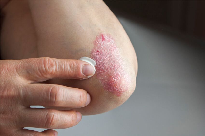
In the first phase of the disease, patients may develop severe fatigue and a vague sense of illness. They may also develop fever and lose weight. In addition, most people with SLE experience joint and muscle pain, leading to severe disability. Skin problems are also common. Patients with SLE commonly develop flat red rashes. These rashes may be associated with calcium deposits or damaged blood vessels. In addition, some patients develop tiny red spots.
TNXB gene
A study has found a possible genetic link between the TNXB gene and systemic lupus erythematosus (SLE). The TNXB gene is involved in the immune system and may be a risk factor for this autoimmune disease. Although this genetic link is not conclusive, it provides insight into the possible mechanisms underlying SLE.
The TNXB gene is a member of the tenascin family of extracellular matrix proteins highly expressed in the skin, tendon sheath, peripheral nerve, and vasculature. Mutations in this gene result in tissue deposition of antibodies, resulting in inflammatory processes and damage to several organs.
HLA-DR4 gene
Recent research on the HLA-DR4 gene and scale disease has found a possible association between this gene and SLE. Specifically, DRB1*03:01 alleles confer a higher risk of SLE than other alleles. The risk is associated with a single amino acid residue in the DR3 peptide-binding groove.
In mouse models of SLE, the HLA-DR4 gene is not implicated in the disease. However, it is implicated in the pathogenesis of the disease in humans. In murine models, this gene has been shown to facilitate the formation of anti-dsDNA antibodies.
TACI-Ig fusion protein
The TACI-Ig fusion protein has demonstrated promise in improving the disease of mice with SLE. It induced a significant decrease in proteinuria and prolonged the survival of SLE mice. Moreover, it prevented the development of the disease in a collagen-induced arthritis mouse model.
TACI-Ig fusion protein inhibits the activity of B-cell lymphocyte stimulator (B-cell activator) and BLyS, two key players in SLE. The fusion protein inhibits the activity of BLyS, a signaling protein in B-cells that triggers autoantibodies' formation.
Hydralazine-induced SLE
Hydralazine-induced SLE is an uncommon manifestation of lupus erythematosus. It is an autoimmune disorder with a variety of symptoms. In addition to the typical erythematous rash, patients may have arthralgia, lethargy, or both. Some patients may experience atypical symptoms such as interstitial lung disease or hypocomplementemia.
This type of SLE presents a skin rash, usually in sun-exposed areas. It may also involve the lower legs. In some cases, the lesions may swell or blister, particularly at the edges of active lesions. The disease is rare and generally begins between eight months and three years after starting the trigger medication. It affects both sexes equally; the mean age at onset is 59 years.


