
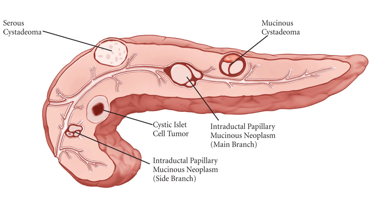
Cysts of the Pancreas Detected With EUS-FNA
Cysts of the pancreas can be detected with CT or EUS-FNA. However, the most accurate and invasive means of diagnosis is through EUS-FNA. It is necessary to understand what this procedure involves. Read on to learn more. Cysts of the pancreas should be treated with specialized therapies, such as surgery or immunosuppression.
Endoscopic ultrasonography with fine-needle aspiration is the most accurate but invasive means of evaluating a cystic pancreas.
EUS is an invasive diagnostic procedure in which a small, flexible tube called an endoscope is inserted into the digestive tract. The endoscope generates high-frequency sound waves and creates a detailed image of the internal organs, lymph nodes, and other nearby organs. Endoscopic ultrasonography is often combined with fine-needle aspiration, a surgical procedure that involves a small needle to sample fluid or tissue from the cyst. The procedure typically lasts no more than an hour.
Among the available diagnostic techniques, endoscopic ultrasonography with fine-needles aspiration is the most invasive but accurate way to evaluate a cystic pancreas. Its accuracy is enhanced by including a mass or nodule within the cyst.
CT abdomen
The differential diagnosis of the cystic pancreas is based on the morphology of the mass. MRI and CT are complementary procedures and can determine whether a pancreatic cyst is malignant or benign. A CT or MRI is usually performed when a CT abdomen shows a solitary pancreatic lesion with a size of three centimeters or less. A thin-section CT can also be helpful.
A CT abdominal examination may show an 8-mm cyst on the pancreas in a patient with chronic right upper quadrant pain. Similarly, a 67-year-old woman presenting with epigastric pain should have her pancreas examined using CT imaging. A CT abdomen with contrast is indicated to determine the cause of her symptoms. A cystic pancreas may appear as a cyst if it has metastasized to another organ.
EUS-FNA
Transgastric EUS-FNA shows a cystic lesion in the tail of the pancreas. Cytology shows reactive pancreatic ducts with ill-defined lobular architecture and no acini. An H and E stained cell-block section reveal cyst debris and foam cells. The diagnosis of the cystic pancreas is based on these findings.
This study focused on 289 patients with suspected PCL. A cystic lesion was identified on cross-sectional imaging, and patients were referred for EUS-FNA. The diagnosis of PCL was presumed after assessing cystic fluid and EUS findings. EUS-FNA and pathological diagnosis are considered the gold standard in determining the cause of PCL. However, this technique is not routinely used in all patients.


